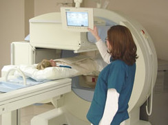
Bone Scan
There are a variety of diagnostic exams your doctor may recommend to determine the cause of your back and/or neck pain, as well as the type of treatment that may be appropriate for you.
 WHAT IS A BONE SCAN?
A bone scan involves intravenously injecting a small quantity of a radiographic marker, into the patient and then running a scanner over the area of concern. The scanner detects the marker, which concentrates in any region exhibiting high bone turnover.
WHY DO I NEED A BONE SCAN?
Your doctor will typically recommend a bone scan when there is suspicion of tumor, arthritis, infection, necrosis or small fractures, i.e., conditions that all result in high bone turnover. A bone scan does not replace diagnostic tests such as an MRI, CAT/CT scan or x-ray imaging, but may provide additional information.
HOW IS A BONE SCAN DONE?
During the exam, your doctor will first give you an injection of the radiographic marker, which contains tiny amounts of radioactive materials called tracers, into a vein in your arm. After two to four hours – the amount of time it takes for the tracers to be absorbed into your bone – you’ll be ready for the actual scan. For the scan, you’ll lie on an exam table while a gamma camera is passed over your body to record the pattern of tracer absorption by your bones. The test may be done in a hospital or outpatient facility, is painless and typically takes about 30 minutes. In some instances, your doctor may order a three-phase bone scan, which involves taking a series of images over a period of time, usually two to four hours.
ARE THERE ANY POTENTIAL RISKS OR COMPLICATIONS?
The risk associated with a bone scan is similar to that of conventional x-ray imaging. There are generally no side effects, and an allergic reaction to the radiographic marker is rare. After the test, you can resume your normal activities.
